Endoscopic Skull Base Surgery: Minimally Invasive Brain Surgery
Skull base tumours are growths at the bottom of the skull, close to the brain, eyes and important nerves. Some can be removed entirely through the nose using a camera and special instruments. This is called endoscopic endonasal surgery, a form of neuroendoscopy. Whether it is suitable depends on the tumour’s position and its relationship with nearby structures.
Hearing that a tumour is “at the skull base” can be worrying. Many people have never heard the term before. Their first questions are usually simple: Where is it? Is it cancer? Will the surgeon need to open the skull?
The answers depend on the individual tumour. Understanding the available brain tumour treatment options can help you prepare for a discussion with your surgeon. Here is what patients and families should understand.
What is the skull base?
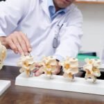
The skull base is the bony floor on which the brain rests. It sits behind the eyes and above the nose and throat.
It has small openings for nerves and blood vessels. These nerves help us see, hear, move our face and swallow. The pituitary gland, which controls several hormones, also sits in this region.
Because so many important structures are close together, even a small growth can sometimes cause problems.
What are skull base tumurs?
A skull base tumour is an abnormal growth in or around this area. It may start in bone, the lining around the brain, the pituitary gland or nearby tissues. Some tumurs grow from the nose or sinuses towards the skull base.
A tumur does not always mean cancer. Some are non-cancerous and grow slowly. Others are cancerous. Even a non-cancerous tumour may need treatment if it presses on a nerve or affects vision or hormone function.
Tumours that may be suitable for an endoscopic approach include:
- Pituitary adenomas: Growths in the pituitary gland that may affect hormones or press on the nerves responsible for vision.
- Selected meningiomas: Tumours arising from the lining around the brain.
- Selected craniopharyngiomas: Growths near the pituitary gland and the nerves involved in vision.
- Clival chordomas: Rare cancers arising in bone near the centre of the skull base.
- Selected nasal and sinus tumours: Growths that involve the front of the skull base.
This list does not mean every tumour of these types can be removed through the nose.
What symptoms can they cause?
Symptoms depend on where the tumour is growing and what it is pressing on. They may include:
- Blurred or double vision.
- Gradual loss of side vision.
- Persistent headaches.
- Changes in smell.
- A blocked nose on one side or repeated nosebleeds.
- Facial numbness.
- Hormone-related changes, such as irregular periods or unexplained breast milk discharge.
These symptoms can have many other causes. They do not, by themselves, confirm a tumour.
Sudden loss of vision, a sudden severe headache or new weakness needs urgent medical assessment.
How is a skull base tumour diagnosed?
The doctor starts with your symptoms, a physical examination and any existing reports.
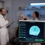
An MRI helps show the tumour and nearby soft tissues. A CT scan may be needed to look at the bone and sinuses. Depending on the location, the doctor may also request vision tests, hormone blood tests or an examination inside the nose.
Some tumours need a biopsy. Others are identified more precisely by testing tissue removed during surgery.
Bring the actual scan images to your consultation, along with the written report. The images help the surgeon decide whether the tumour can be reached safely through the nose.
How is the tumor removed endoscopically?
The operation is performed under general anaesthesia, so you are asleep.
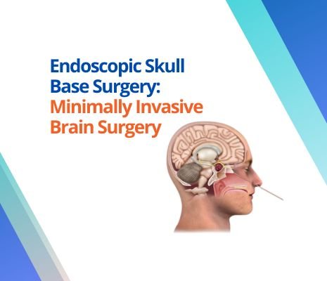
The surgeon passes an endoscope—a thin instrument with a light and camera—through the nostrils. It shows a close view of the operating area on a screen. Fine surgical instruments are passed alongside it.
The team creates a carefully planned route through the nose and sinuses. A small amount of bone may need to be removed to reach the tumour. The surgeon then separates the growth from nearby tissues and removes as much as is safe.
If the protective covering around the brain is opened, the team repairs the opening. Tissue from inside the nose may be used to help seal it and prevent leakage of the fluid surrounding the brain.
An ENT surgeon and a neurosurgeon often work together during this operation.
What does “purely endoscopic removal” mean?
It means the tumour is approached and removed using endoscopic instruments, without an additional open operation through the skull.
For suitable tumours, the entire procedure can be performed through the nose, without an external cut on the face or scalp.
However, purely endoscopic surgery does not automatically mean complete tumour removal. If part of the tumour is firmly attached to an important nerve or blood vessel, leaving a small portion may be safer. Further treatment or monitoring may then be needed.
Can every skull base tumour be removed this way?
No. The nose provides a useful route to certain areas, particularly near the centre of the skull base. Tumours extending far to the side or surrounding important blood vessels may need a different approach.
Some patients need open brain surgery or a combination of approaches. Others may benefit from medicines, radiotherapy or observation rather than immediate surgery.
The choice should protect vision, nerve function and long-term health. The scan findings matter more than choosing a particular technique. If you are unsure about a recommended operation, a second opinion for brain surgery can help you understand the reasons for the proposed approach.
What are the benefits and risks?
For carefully selected patients, the endoscopic route can avoid an external scar and reduce the need to move brain tissue to reach the tumour.
It remains a major operation. Risks include bleeding, infection, leakage of the fluid around the brain, and injury to nerves or blood vessels. Some patients may develop changes in vision, smell or hormone function.
Your surgeon should explain the risks for your particular tumour, including the possibility of further treatment.
What is recovery like?
A blocked nose, tiredness and some discomfort can occur after surgery. Recovery varies with the tumour, the extent of surgery and whether a skull base repair was needed.
The team will explain when you can return to work and how long to avoid heavy lifting, straining and blowing your nose.
Follow-up may include nasal checks, MRI scans, vision assessments and hormone tests. Clear watery drainage from the nose, fever, a worsening severe headache or new vision changes after surgery needs prompt medical attention.
Seeking skull base tumor surgery in Pune
If your scan shows a skull base tumour, a consultation should help you understand what it is, whether treatment is needed and which approach is safest.
For assessment with Dr. Vishal Bhasme, a neurosurgeon in Pune, bring your MRI or CT images, reports and current medicine list. Patients seeking care at Lopmudra Meera Hospital, Camp–Swargate, can book a specialist consultation.
Ask: “Can my tumour be removed entirely through the nose, and what might prevent complete removal?” It is a practical question that helps you understand the plan.
Frequently asked questions
Is a skull base tumour the same as brain cancer?
No. It describes the location of a growth. Some skull base tumours are non-cancerous, while others are cancerous.
Does endoscopic surgery leave a visible scar?
A purely endonasal operation usually does not leave an external scar on the face or scalp. Additional tissue harvesting, if required, may involve another incision.
Is surgery always needed?
No. Treatment depends on the tumour type, growth, symptoms and scan findings. Some tumours can be monitored; some respond to medicines or radiotherapy.
Can a tumor return after removal?
Yes, some can grow again. Follow-up scans are important even when the surgeon believes the tumour has been fully removed.
Will I need radiotherapy after surgery?
Some patients do. The decision depends on the tissue diagnosis, any remaining tumour and the risk of regrowth.
This blog provides general information. It is not medical advice and does not replace an individual consultation.
Publishing notes
- Use the article title as the page H1; retain the smaller heading appearance where preferred.
- Eight contextual internal links have been embedded in the article, pointing to existing pages on Dr. Vishal Bhasme’s website.
- Link relevant older brain tumour and pituitary tumour articles back to this article.
- Suggested image alt text: “Illustration showing an endoscopic route through the nose to a skull base tumour.”
- Add MedicalWebPage or Article schema and verified Physician information. Include FAQPage markup only for the FAQs visible on the page. Do not mark the draft as medically reviewed before approval.
Medical references
- Johns Hopkins Medicine: https://www.hopkinsmedicine.org/health/conditions-and-diseases/brain-tumor/skull-base-tumors
- Johns Hopkins Medicine: https://www.hopkinsmedicine.org/health/treatment-tests-and-therapies/endoscopic-endonasal-surgery
- Johns Hopkins Medicine: https://www.hopkinsmedicine.org/health/treatment-tests-and-therapies/skull-base-surgery
- UCLA Health: https://www.uclahealth.org/medical-services/neurosurgery/pituitary-skull-base-tumor/treatment-options/endoscopic-skull-base-surgery
- UPMC: https://www.upmc.com/services/neurosurgery/brain/treatments/endoscopic-endonasal-approach/conditions