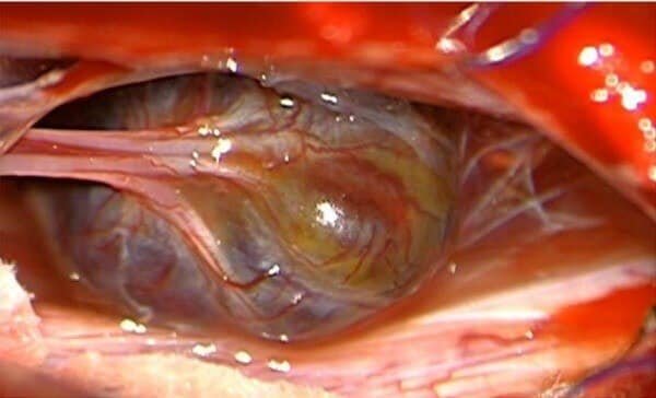
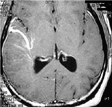
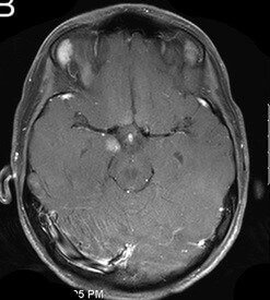
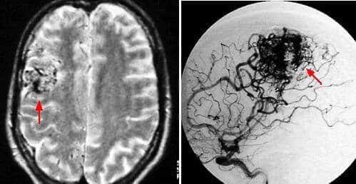
These are abnormal tuft of arteries and veins in the brain. They are present since birth. Later with time they increase in size pushing the brain parenchyma leading to perilesional fibrosis. Once they bleed, they result in increase in the mass effect and cause symptoms, depending on their size and location.
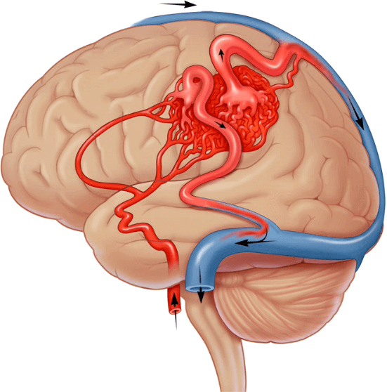
AVMs are classified and graded according to a scoring system devised by Spetzler-Martin. Accordingly, the treatment is offered to the patient. Spetzler-Martin system classifies the AVM based on the size (<3 cm, 3-6cms, >6 cm), involvement of any important area of the brain (eloquent / non-eloquent) and the venous drainage (superficial / deep).
Low grade AVMs are associated with less chances of bleeding comparatively with the high grade AVMs.
- Lower grade (I & II) is amenable to open surgical removal or endovascular treatment
- Medium grade (III) requires both endovascular treatment along with radiation/open surgical removal
- High grade (IV & V) very difficult to treat.
When AVM bleeds, depending on the characteristics, treatment is sometimes offered to prevent more brain damage from additional future bleeding. If AVM is found before it was bled, the treatment can be observation or definitive. The treatment methods include radiation, open microsurgical procedure and/or endovascular treatment. It is always important to meet with your concerned neurosurgeon and discuss the pros and cons of each procedure and the best available option.
Other vascular malformations include cavernomas, venous angiomas and capillary telangiectasia. These have very less propensity to bleed. So, there treatment mainly includes observation. Only sometimes very few cavernomas require treatment.