Brain Tumor Surgery in Pune
Brain Tumor Surgeon in Pune
Surgery is the main treatment for most primary brain tumors. Dr. Vishal Bhasme has done numerous successful brain tumor surgery in Pune. Brain tumors can be classified in many ways. The most common among them are metastatic tumors. These tumors spread to the brain from another organ in the body. The most common tumor that starts in the brain is called an astrocytoma.
The symptoms depend upon the size and the location of tumor. The commonest symptoms include the focal neurological signs like language/speech problems, motor weakness, altered sensations, vision problems, change in personality, seizures. Sometimes, it may cause increase intracranial pressure which leads to headaches, vomiting and loss of consciousness.
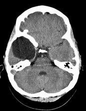
Diagnosis is usually done by radio-imaging studies like CT and MRI brain.
Secondary Brain Tumors: These are tumors from some other organ of the body and infiltrate into the blood vessels. Tumor cells from there migrate into the brain and start growing there, forming the secondary brain tumor or metastatic tumor. Common malignancies which migrate to the brain includes lung, breast, melanomas (skin) and renal cancer. These are the most common tumors of brain. Around 25% of patient dying from cancer have brain metastasis.
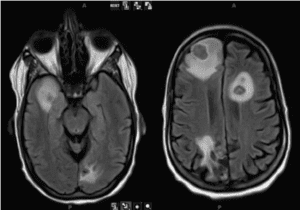
Treatment of such tumor is very complicated. Most of the time it is a stage 4 disease, so patients are treated by chemotherapy and radiotherapy. Only in few patients, when there is only one or few lesions and the overall prognosis is reasonable, surgery is advisable. Patients with brain metastases often are more likely to die from their brain tumor than from the cancer in the rest of their body.
To treat such cases a multidisciplinary team approach is adopted which includes a neurosurgeon, medical oncologist & radiation oncologist to give the best outcome to the patient.
Primary Brain Tumors– These arise from the brain parenchyma or the surrounding structures.
They are further classified into:
- Cancerous
- Non-Cancerous (Benign)
Glioblastoma Multiforme
Cancerous brain tumors are those which cannot be completely excised and have a high chance or recurrence. Most common among them is Glioblastoma Multiforme (GBM). It is a type of grade-4 astrocytoma. Usually occur in elderly population, but may present at any age. It may spontaneously mutate from a low-grade astrocytoma. There is no conclusive evidence for the causes of such tumors, but the risk factor includes ionizing radiation and genetic predisposition. Since last few years tremendous research has happened over some genetic factor linked with GBM. Tumor tissue is sent to look for certain genetic changes which have been linked with better survival.
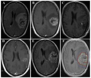
Median survival rate for GBM after maximum treatment which includes surgery, chemo and radiotherapy is around 15 months.
Surgery remains the initial part of the treatment for most of the brain tumors, including GBM. It infiltrates the brain like the tentacles of octopus and rarely has defined margins, so it is quite impossible to remove the tumor completely. Instead, the bulk of the tumor is removed surgically to reduce the tumor load, followed chemotherapy and radiation. Whenever tumor is involving the important part of brain, only a small biopsy is taken and rest of the treatment is done using chemotherapy and radiation. This is done mainly, not to impart any new deficit and maintaining the quality of life.
Nowadays, many new surgical tools are available to remove such tumors with maximum safety. We have high end microscopes, neuro-navigation (GPS) system due to which such surgeries have become very safe. We perform surgeries in some patients when he is awake which allows us to check his speech, movements and sensation intra operatively. Minimally invasive brain surgery is a new branch now, where we use cameras, called an endoscope or a small tubular retractor system to reach the deeper brain tumors and remove the maximum tumor tissue without damaging the surrounding normal brain and hence reducing the collateral damage.
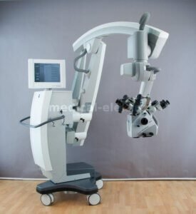
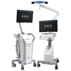
Recently, the chemotherapy regimens have also improved and target certain genetic receptors of the tumor.
Immunotherapy, cancer vaccines and few viruses are being researched for exploring new treatment strategies, which are mainly reserved for recurrent disease.
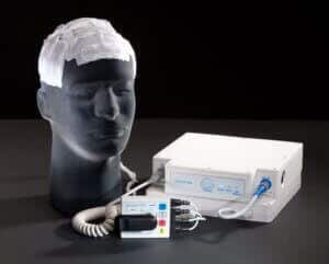
OPTUNE, an electromagnetic therapy which involves wearing a cap containing electrodes on the scalp is a new treatment strategy to treat GBM.
Other Astrocytomas
World Health organization (WHO) histopathologically has developed the grading system for astrocytomas in which the grade-1 being the least cancerous (benign) and grade-4 the most cancerous.
GRADE-3
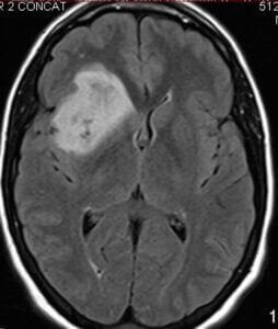
These are anaplastic astrocytomas and are treated on the similar lines of grade-4.
GRADE-2
These includes
- Pleomorphic xanthoastrocytoma
- Low grade diffuse astrocytoma
- Oligodendroglioma
- Ependymoma
- Central Neurocytoma
- Certain Meningiomas
These tumors are treated on case-to-case basis, with few smaller asymptomatic lesions treated conservatively with repeated Mri Scans. Whenever there is a substantial increase in the size of the tumor or edema, surgery is done followed by chemo and radiotherapy.

GRADE-1
These are benign tumors, and can be observed like grade-2 lesions but if there is any seizure activity or neurological deficit, they are surgically removed. These include
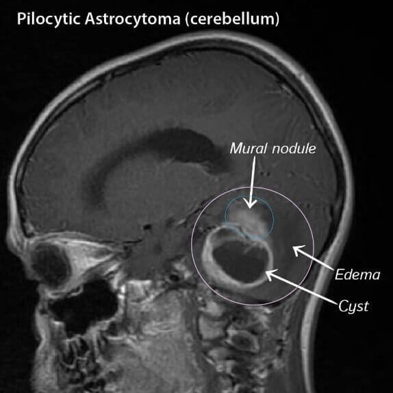
- Pilocytic Astrocytoma
- Sub-ependymal giant cell astrocytoma (SEGA)
- Sub ependymoma
- Ganglioglioma
- DNET
- Pineocytoma
- Choroid
- Schwannoma
- Neurofibroma
- Meningioma
- Pituitary Adenoma
- Craniopharyngioma
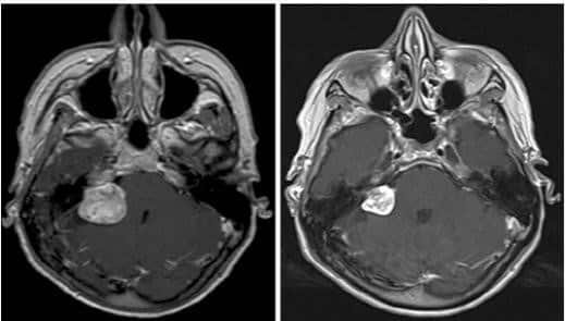
Meningiomas are again one of the commonest brain tumors which require surgery. They mainly arise from the meninges which are the covering of the brain below the skull bone. They are very slow growing tumors and often become symptomatic once they become very big and start pushing the surrounding structures.

Other tumors:
These are few tumors which are quite benign and are present by birth, and gradually start increasing in size very slowing and become symptomatic once they start pushing the surrounding structures
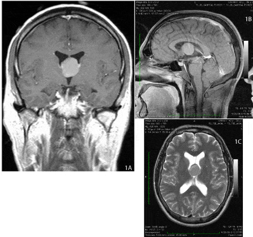
Colloid Cyst: It is a congenital cyst which grows which resides in a natural fluid filled cavity called the 3rd ventricle. When it grows, it starts obstructing the CSF flow of the lateral ventricles resulting in drop attacks, sometimes can be life threatening. Urgent surgery is required in such cases. It is removed using a microscopic or an endoscopic procedure. The size of the incision in endoscopic procedure is just 2 cms.
Epidermoid/Dermoid Cyst: These are inclusion cysts which are present by birth and enlarge with time producing pressure effect on the surrounding structures and need surgery. These cysts mainly occur in the midline at the posterior fossa region.

Arachnoid Cysts: These are not actually tumors, but is the fluid filled cavity lined by the arachnoid membrane. These are also congenital lesions which very rarely requires a surgical treatment. These are very commonly found as an incidental finding and are treated conservatively. For those patients require surgical treatment, endoscopic drainage of the cyst into a cistern space is done.
